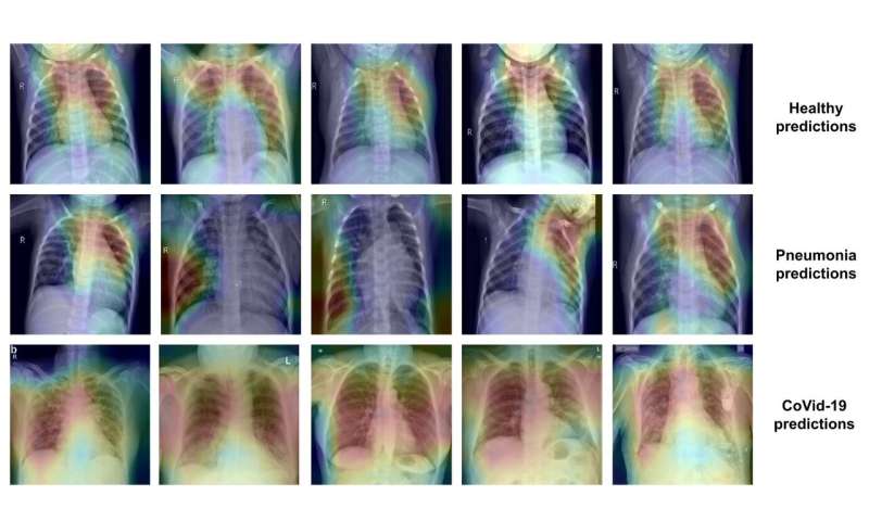
A research team from Valencia’s Polytechnic University (UPV), from the CVBLab, has developed a predictive artificial intelligence model that can tell the difference between healthy patients, those who are ill with pneumonia and those who have COVID-19, from chest X-rays.
According to Valery Naranjo, professor at the UPV and CVBLab director, the proposed model has proven to have great discriminatory capabilities in the first experiments, reaching an average success rate of 92% when differentiating between the different types of patients. “The algorithm behaves even better when predicting cases of coronavirus; its success rate is slightly better compared to the other cases: it has a success rate of 97% when determining whether an X-ray is from a COVID patient,” notes Valery Naranjo.
The CVBLab research group of the UPV has comprehensive experience in the field of artificial intelligence and their specialty is developing computer vision algorithms applied to biomedical images. “This is why we have put our knowledge to the service of the fight against this pandemic,” concludes Julio Silva, biomedical engineer and also a member of the CVBLab of the UPV.
To develop the prediction model, the CVBLab engineers have applied classification and segmentation techniques based on deep learning algorithms on a large number of X-ray images. In this sense, Valery Naranjo explains that there are many more X-rays from healthy people and patients with other pneumonias than with COVID-19,” due to how recent it is and because many databases are not open source, which represents an added difficulty. The model we have developed solves this class—patient—unbalance and makes it possible to offer reliable and robust results.”
The CVBLab group already has an initial version of the computer platform that integrates the prediction model, so it is possible to load a chest X-ray and instantly predict if it is an image of a healthy person, a patient with pneumonia or with coronavirus.
Innovative design
The artificial intelligence model of the CVBLab-UPV displays key novelties in the design of the neural network architecture. In particular, it is based on knowledge transfer techniques combined with other residual convolutional blocks that act in parallel to extract characteristics from chest X-rays.
“This new design, adapted to the type of image being studied, has made it possible to obtain initial sensitivity and specificity results of 97%,” adds Gabriel García, biomedical engineer and researcher from the CVLab of the UPV.
CBIR system
At the same time, the researchers are developing a new content-based image retrieval (CBIR) system based on generative neural networks. The idea of this system is that, upon receiving a new X-ray image, as well as obtaining a prediction on the diagnosis, the most similar prior cases from a large and constantly growing database will be provided. “The affected lung areas from the archives of the most similar cases are shown with a very intuitive heat map for the expert personnel that uses it. Thus, the doctor has more data in order to make a decision. It is like when they look for something in an atlas, but automatically,” says Adrián Colomer, doctor in telecommunication and researcher from the CVBLab of the UPV.
To create their models, the CVBLab researchers of the UPV have compiled public databases from different institutions, and have normalized them in a common framework, which makes it possible to train and test their models.
Source: Read Full Article
