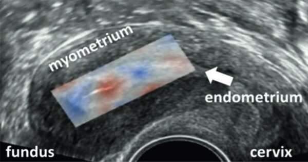
In groundbreaking work for women with fertility problems, Eindhoven University of Technology (TU/e) and the Catharina Hospital have developed a new method that allows for simple and objective measurements of uterine contractions. Measuring uterine ‘waves’ correctly is important, as they play a key role in the implantation of an embryo in the uterus. However, the methods available so far to measure them were not reliable. The new tool, based on a technique used by cardiologists to measure heart motions, gives gynecologists new insights into uterine contractions, and will increase our understanding of their impact on the implantation of an embryo in IVF.
Every uterus exhibits ‘uterine waves’ during a monthly cycle: small undulating contractions in the uterus that you don’t feel, but can see on, for example, ultrasound images. These waves change direction and intensity during the cycle depending on hormones. During IVF treatment, uterine contractions provide nutrients, prevent the embryo from being expelled from the cavity, and contribute to the positioning of the embryo before implantation.
Increasing the chance of pregnancy
“Understanding these ‘waves’ is crucial to increase the success of IVF,” says researcher Celine Blank, who will receive her Ph.D. from TU/e and Ghent University on Friday, February 26. “Now we have this new tool, we will be able to get to know uterine contractions better. They can serve as a predictive factor in fertility treatments. The success rate for women undergoing fertility treatment is now around 30 percent per IVF cycle. That rate really needs to go up! Treating a shortage or surplus of waves with medication, for example, can potentially increase the chance of pregnancy.”
The technique used to measure uterine waves was copied from cardiologists. Dick Schoot, gynecologist at Catharina Hospital and Blank’s supervisor: “Cardiologists use the Speckle Tracking technique to measure heart motions, which, like the uterus, is also a muscle. That method was adapted for the uterus and then tested extensively. First in a controlled environment on uteruses outside the body, and later in healthy volunteers.”
“Slowly but surely, we understand better which mechanical movements of the uterus play a role in pain, irregular bleeding and getting pregnant. But we are still at the beginning of this promising technique,” says Schoot.
Less stress for women
He continues, “We combine this examination with electricity measurements of the uterus. Such an examination is not at all stressful for the woman. For the measurements, stickers are placed on the abdomen to measure the contractions. You can compare it to a heart monitor (ECG).”
Source: Read Full Article
