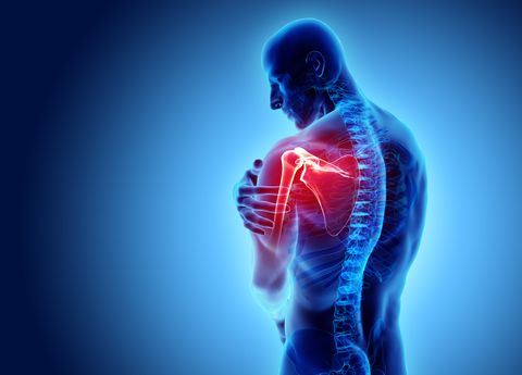
Put down that dumbbell and listen up: Muscles aren’t everything.
“It’s your joints that make the whole body tick,” says Douglas Comeau, D.O., director of the Boston University Sports Medicine Fellowship. But like any mechanical system, they’re prone to wear and tear. And without well-functioning joints, it’s challenging to add muscle, shed fat, or get anything done around the house. To maintain them, you need to understand how they work and the threats they face.
There are three important types of joints:
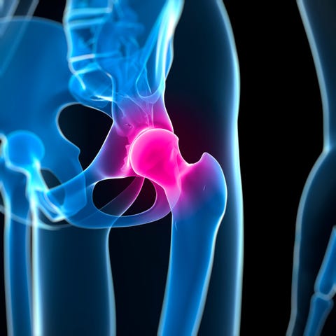
Ball-and-socket
Getty ImagesSCIEPRO
The rounded, ball-like end of one bone fits into the concave surface of another. The design offers superior range of motion, but stability can vary—high in a deep socket, low in a shallow one. Surrounding tendons and ligaments help keep bones in place.
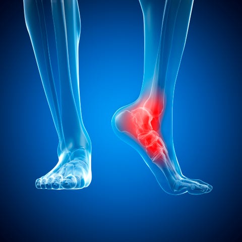
Hinge
Getty ImagesSEBASTIAN KAULITZKI
Adjoining bones flex toward or extend away from each other in a swinging motion along an axis. Ultrasmooth articular cartilage between the bones reduces friction as they move against each other.
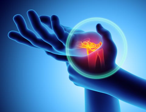
Ellipsoid
Getty Imagesyodiyim
In a modified ball-and-socket, the rounded end of one bone (or multiple bones) moves against a shallow, elliptical cavity in another, allowing a wide range of movement.
Here’s how to keep your six major joints jumpin’.
THE SHOULDER

Bryan Christie Design
Type of Joint: Ball-and-socket
Shoulders have an exceptional 360-degree range of motion but a shallow socket and relatively loose ligaments. “What you gain in mobility you lose in stability,” says Brian Sennett, M.D., chief of sports medicine and vice-chair of orthopedic surgery at the University of Pennsylvania Health System.
Top Threat: Labral tear
What it is: Damage to the shoulder labrum, a rim of fibrous cartilage that gives the shoulder socket its cuplike shape. A labral tear makes it harder for the ball to stay seated in the socket, so dislocation often follows.
Cause: Usually trauma—breaking a fall with an outstretched arm or dislocating a shoulder in an accident—but overuse from throwing or lifting can fray the labrum, too.
Treatment: Rehab can strengthen muscles and shore up supporting tendons to stabilize the shoulder. If it doesn’t or there’s danger of dislocation, surgery is usually needed to trim frayed or loose labral tissue or reattach the labrum to the socket.
Defense: Do this simple exercise: Stand to the right of a resistance band fastened at waist height. Holding the end in your right hand, lock your right elbow to your side and slowly rotate your arm outward, pausing in the fully rotated position. Do 15 reps, then stand to the left of the band and rotate inward against resistance. Repeat with your left arm.
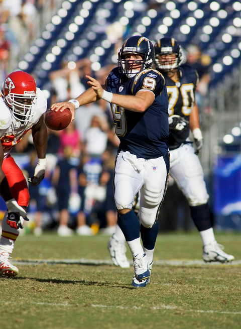
Future-Proof Your Shoulders: Overdoing overhead motions like swimming, throwing a baseball, swinging a racket, or even painting walls can result in impingement on your rotator cuff, a group of muscles and tendons that cover the head of the upper arm bone and hold the joint in place. This can cause the cuff to tear, especially as tendons become weaker and stiffer with age.
To prevent it, do this exercise: While standing, lift a 1- or 2-pound dumbbell to the side and about 30 degrees forward with arm straight and thumb pointed down, like you’re pouring a beer. Do 15 reps per side. According to orthopedic surgeon Nicholas DiNubile, M.D., this isolates and strengthens the supraspinatus muscles to support the tendon most often damaged.
Watch Out!
A popular impingement surgery called subacromial decompression, which smooths bone spurs on the acromion, may not accomplish much, according to a recent study in The Lancet. Patients in 32 UK hospitals who got the arthroscopic procedure didn’t do any better than a control group that had a sham surgery in which doctors scoped the shoulder but didn’t fix anything. Both groups did only slightly better than people who received no treatment, leading researchers to suggest that other treatments, such as rehab, painkillers, and steroid injections, may be more beneficial.
THE HIP

Bryan Christie Design
Type of Joint: Ball-and-Socket
Hips have half the mobility of shoulders but a much deeper socket, which makes the joint highly stable—essential for bearing weight, walking, running, jumping, and drunk dancing at weddings.
Top Threat: Hip labral tear
What it is: An injury of the hip labrum, a gasket-like cartilage ring at the rim of the hip socket that helps hold the ball of the thighbone in place and seals in fluid. Besides causing pain, labral tears raise the risk of hip osteoarthritis.
Cause: Usually repetitive motion, such as from long-distance cycling, or collisions in sports. “We see it a lot in cutting sports like soccer and hockey,” Dr. Comeau says. Tennis players are also prone to hip trouble. Former top-ranked pro Andy Murray finally had surgery in January to ease hip issues that had plagued him for years. Abnormal hip anatomy can also contribute.
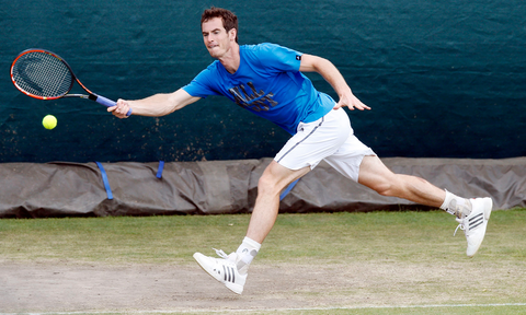
Treatment: Physical therapy can help identify and compensate for quirks in your gait or anatomy that may stress the hip, and stretch and strengthen hip-supporting muscles. If these approaches don’t work, a surgeon can use an arthroscope to trim frayed cartilage and reattach the labrum to the socket.
Defense: Vary your sports and workouts from day to day to avoid stressing the hip in the same way and to help joints recover.
Future-Proof Your Hips: A previous hip injury or normal aging can erode the articular cartilage that lines the hip’s ball and socket and lead to osteoarthritis. As cartilage diminishes and the space between the bones closes, damaged bones may grow out and form spurs that can add to pain and limit movement. To keep your hips young, do bridges, planks, and lunges to strengthen your gluteus, lower back, and hip-flexor muscles, which support and stabilize the hips. Don’t do lunges while holding weights, though, to avoid undue stress.
Watch Out!
Metal-on-metal hip implants haven’t worked out as well as expected. The ball-and-socket combo promised to be exceptionally durable, but the FDA warns that they carry “unique risks,” including the release of metal particles that may damage surrounding bone and/or tissue. Consider alternative bearing surfaces such as ceramic on cross-linked polyethylene instead.
THE ANKLE
Bryan Christie Design
Type of Joint: Hinge
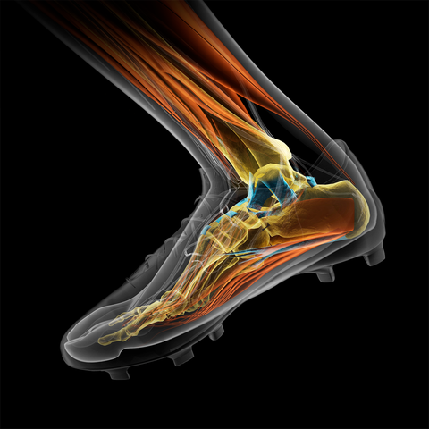
The ankle is highly stable in a neutral standing position. But in a downward-flexed, on-your-toes position, the joint depends more on support from injury-prone ligaments and tendons.
Top Threat: Sprain
What it is: A tear in one of the ligaments — usually on the outside of the ankle — that supports the joint. Severe sprains that leave the ankle unstable may eventually damage the joint’s bones and cartilage.
Cause: Stretching the ligament beyond its limits, usually by rolling the foot as you walk or run on an uneven surface, making a cutting move, or stepping on someone’s foot. Just a month ahead of Portugal’s 2018 World Cup bid, star forward Cristiano Ronaldo sprained his ankle and had to leave the game. “Outside of sports, the most common scenario for men is rolling the foot while going down a step,” says Rocco Monto, M.D., spokesman for the American Academy of Orthopaedic Surgeons.
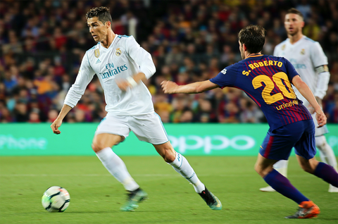
Treatment: Rest and compress the ankle, possibly with help from a brace. Rehab will build supporting muscles and increase balance with exercises like standing on one foot with eyes closed— important for preventing repeat sprains due to ankle instability. Surgery to reconstruct the ligament and brace the ankle is rarely needed.
Defense: Build calf muscles with exercises like calf raises to increase support around the ankle and improve balance. “They’re like stirrups that hold the ankle in,” says Dr. Monto.
Watch Out!
It’s important to keep broken anklebones immobile, but that doesn’t mean you have to be. “You want the joint to bear weight because that generates electrical fields that stimulate the bone to heal,” Dr. Monto says. Figure on an active recovery, not a restful one.
ELBOW
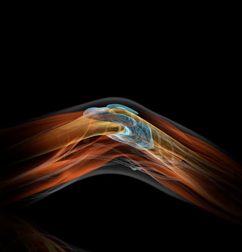
Bryan Christie Design
Type of Joint: Hinge
Tendons attaching bones to muscles in both the upper and lower arm come together in the elbow area. Ligaments hold bones tightly in place and stabilize the joint.
Top Threat: Olecranon fracture
What it is: A crack or break in the elbow’s tip; this can result in an open fracture in which bone sticks through skin and can cause infection.
Cause: Usually direct impact with a hard object, such as getting hit by a baseball or whacking your elbow against a doorframe. Falling on an outstretched arm can also stress the joint enough to separate bone, as can repeatedly poking your elbow into the ribs of someone with zero sense of humor.
Treatment: If pieces of bone aren’t out of place, splinting for about six weeks should allow the fracture to heal. More complex fractures require surgery to realign bone fragments. A graft can fill in bone that’s been lost or destroyed.
Defense: Wear elbow pads in sports such as mountain biking and snowboarding, which have a higher risk of falls. And just in case you do go down, practice the tuck and roll. On a soft surface, crouch down, bend forward, tuck your head, and roll onto one shoulder. Try to curl into a ball as you do so, using your arms for guidance rather than as shock absorbers.
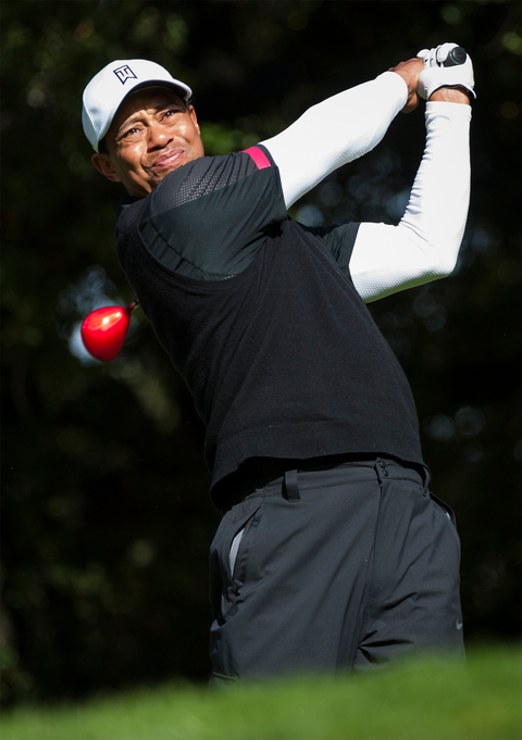
Future-Proof Your Elbows: If you’re a golfer or tennis player, take lessons to learn how to swing a club or racket using your core and whole body, not just your arms. “A poor swing puts chronic strain on tendons, causing overuse damage that can be difficult to heal,” says Darryl D’Lima, M.D., Ph.D., of the Scripps Clinic in La Jolla, California.
This can result in epicondylitis, a painful inflammation of tendons that connect forearm muscles to bony protrusions called epicondyles on the outside and inside of the elbow. You know outside (lateral) epicondylitis as tennis elbow and inside (medial) epicondylitis as golfer’s elbow, but both can occur in either sport.
Bonus tip: Ease into your game. “Massage your forearm muscles at red lights on the drive over,” says Dr. DiNubile. “That stimulates blood flow to the area around the joint.” Hit the first few balls gently, working up a light sweat to limber up muscles and ligaments before taking more vigorous strokes.
Watch Out!
“If your tennis racket isn’t strung correctly, it will transmit more force up the forearm to the elbow,” says Dr. D’Lima. A recent study in Shoulder & Elbow found that lower string tension placed less of a load on the elbow, potentially reducing the risk of lateral epicondylitis.
THE WRIST
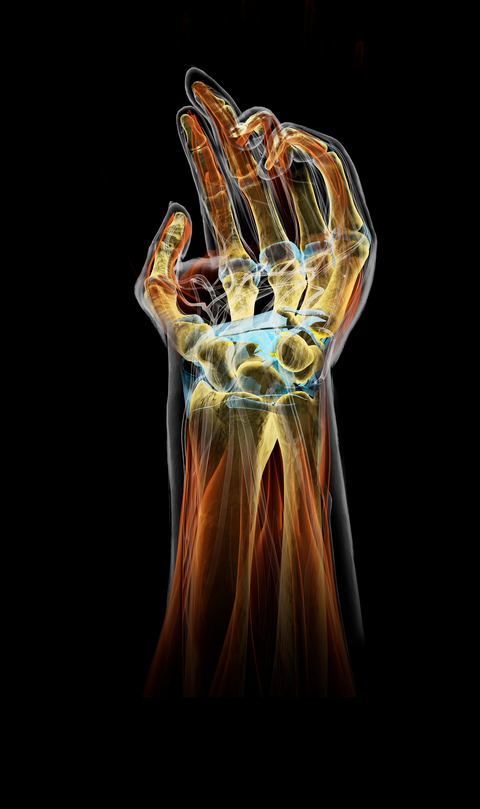
Bryan Christie Design
Type of Joint: Ellipsoid
The wrist’s collection of bones, ligaments, tendons, and cartilage forms the body’s most complex joint. Because it’s not weight-bearing, it’ll likely provide problem-free mobility for a lifetime—unless you injure it.
Top Threat: Scaphoid fracture
What it is: A break in the scaphoid bone, one of eight small carpal bones in the wrist. (Stick out your thumb hitchhiker style: The scaphoid is under that little divot at the base of your thumb.) Scaphoid breaks account for about 70 percent of carpal fractures.
Cause: Usually falling palm down on an outstretched hand—an injury that often occurs in young men.
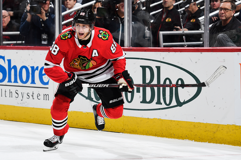
Treatment: A cast or splint that immobilizes the thumb for about six weeks can treat most fractures, especially close to the thumb, where there’s good blood circulation. If bones are displaced, aren’t healing, or show signs of decay due to poor blood supply, you may need surgery to align them and hold them in place with screws, pins, or wires. A year after scoring the winning Stanley Cup goal, Patrick Kane of the Chicago Blackhawks had surgery to repair a scaphoid fracture. He’s since won two more Cups with the ’Hawks.
Defense: If you don’t want to quit the sports most likely to break wrists—football, soccer, hockey, skiing, snowboarding, in-line skating—wear wrist guards and/or learn to tuck and roll.
Future-Proof Your Wrists: A triangular fibrocartilage complex (TFCC) tear entails damage to the cartilage on the pinkie-finger side of the wrist that supports and cushions the carpal bones. TFCC tears cause pain near the pinkie, especially when bending the wrist from side to side. As with a scaphoid fracture, it can stem from falling on an outstretched hand (or violently twisting the wrist).
Another cause: the sudden binding of a power-drill bit that spins the drill while it’s in your grip. Protect yourself by upgrading to a drill with a torque grip that lets you grab two handles for better control, or kickback control that cuts power when the drill starts rotating.
Watch Out!
If an X-ray doesn’t show a scaphoid fracture immediately, wait. Small breaks often don’t appear until 10 to 14 days after injury, when poor blood supply causes bone decay that’s more visible on a scan. Wrist still hurting after two weeks? X-ray it again.
THE KNEE
Bryan Christie Design
Type of Joint: Hinge
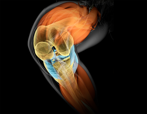
Four main ligaments bind the thighbone (femur) with the shinbone (tibia) and kneecap (patella). Knees are versatile and strong but vulnerable to a variety of breakdowns.
Top Threat: Anterior cruciate ligament (ACL) tear
What it is: A sprain of the front-most of two ligaments that cross each other (hence cruciate) inside the knee. A mild sprain may stretch the ligament without affecting stability, but more severe sprains can partially or completely tear it, leaving the joint loose and likely to further damage the knee’s cartilage.
Cause: Excessive force from pivoting in a pickup basketball game, jumping to spike a volleyball, or colliding with something much larger than you. That’s why ACL tears are common among athletic younger men.
Treatment: Surgery is usually needed, especially if you want to play sports again. Most ACLs can’t be stitched together, so the surgeon usually takes a graft of your patella or hamstring tendon (or a cadaver tendon) and uses it as scaffolding to reconstruct the ligament.
Defense: Stronger leg muscles make knee joints more stable. To strengthen yours, do these three exercises in a series: walking lunges (3 sets of 10 reps), Russian hamstrings (kneel on the floor with a partner holding your ankles, then lean forward with a straight back for 3 sets of 10 reps), and single toe raises (30 reps each leg).
For a complete knee-protection plan, visit health.usf.edu and search for the PEP program from the Santa Monica Sports Medicine Research Foundation. It provides a 15- to 20-minute regimen that includes the leg exercises outlined here, plus warmups, stretches, strengthening exercises, and plyometrics that significantly reduce ACL injuries in athletes.
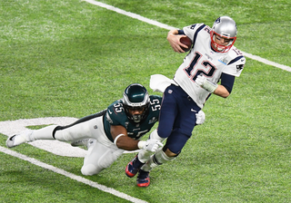
Future-Proof Your Knees: The forceful movements that cause ACL tears can also damage the meniscus, a rubbery cartilage wedge that provides cushioning inside the knee. Normal aging also weakens the cartilage, so just getting up from a crouch (or couch!) can tear an older knee. “We see it a lot in baseball catchers, plumbers, and carpet layers—anybody who torques their knees for a living,” says Dr. DiNubile. The aforementioned leg-strengthening PEP program for protecting the ACL is a good defense here, too.
Watch Out!
Unless their meniscus tears are due to trauma such as a sports injury or fall, people who undergo a partial meniscectomy do no better than those receiving sham surgery or physical therapy, according to recent studies. “If you’re over 50, when a torn meniscus is probably degenerative, you’re likely better off with physical therapy and maybe cortisone injections,” Dr. Sennett says.
DIY REPAIR
Getty ImagesWestend61
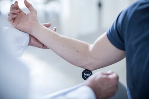
The best joint repairs are the body’s own. “Tissues naturally replicate and, through healing, get better over time,” says Darryl D’Lima, M.D., Ph.D. That’s why researchers are studying ways to boost the body’s self-repair with techniques like these:
Platelet-rich plasma
Platelets in blood plasma contain proteins called growth factors that promote healing. In PRP therapy, your doc draws your blood, spins it to separate and concentrate the platelets, and injects the preparation into a joint, hoping to help the healing process. Studies suggest it’s most effective for chronic tendon injuries, especially tennis elbow, but research hasn’t conclusively shown it’s better than existing treatments.
Stem cells
Stem cells harvested from bone marrow (new research is looking into extracting them from fat) have the potential to grow into other types of cells. Once programmed in the lab, they’re put into a joint to grow new tissues like cartilage and ligament. The therapy has shown promise in animal studies, and some clinics offer it for osteoarthritis. “But the jury is still out,” says Dr. D’Lima.
Cartilage implants
The FDA recently approved a procedure called autologous chondrocyte implantation (ACI) that helps heal damaged cartilage in people without osteoarthritis. A surgeon first extracts one or two small samples of cartilage from your knee in the non-weight-bearing zones. These are processed in a lab, and the cells are placed back into the knee joint during a second operation. The ACI procedure existed before, but cells are now grown on a membrane that’s glued to the damaged cartilage, providing a scaffold that accelerates repair. “It can be helpful for young and middle-aged patients with isolated cartilage defects,” says Kevin D. Plancher, M.D., of Albert Einstein College of Medicine.
Enhanced ACL repair
In a new technique, a scaffold loaded with a patient’s blood is placed between the ends of a torn ACL before they’re sutured. Called bridge-enhanced ACL repair, or BEAR, it stimulates healing in the ligament and doesn’t require a graft from elsewhere in the body. “Good preliminary results,” says Brian Sennett, M.D.
Source: Read Full Article
