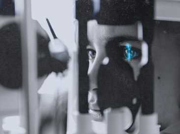
- A noninvasive eye scan makes it possible to assess the difference between retinal age and chronological age.
- New research suggests that this retinal age gap may be linked to mortality risk from some causes.
- The study found no association with deaths from cardiovascular disease or cancer.
- The researchers suggest that the retinal age gap could serve as a screening tool.
Anyone who undergoes routine eye tests is likely to have undergone retinal imaging. A specialized camera takes images to check the health of the tiny blood vessels in the retina, the light-sensitive layer at the back of the eye.
Previous research has shown that the health of the retina may be a biomarker of conditions such as diabetes and cardiovascular disease.
Now, a study in the British Journal of Ophthalmology suggests that healthcare professionals could use retinal photography to predict mortality risk. In this study, researchers from China, Australia, and Germany used deep learning to assess retinal age based on images of the back surface — called the fundus — of the eye.
Dr. Howard R. Krauss, a surgical neuro-ophthalmologist and clinical professor of ophthalmology and neurosurgery at Saint John’s Cancer Institute at Providence Saint John’s Health Center in Santa Monica, CA, told Medical News Today:
“Zhu [and colleagues] have demonstrated that the application of artificial intelligence coupled to deep learning to discern an ‘apparent’ retinal age, when compared to the true age, is an indicator of risk of mortality.”
– Dr. Howard R. Krauss
Retinal age gap
The study investigated the “retinal age gap” in 35,913 people, using data from the UK Biobank. The retinal age gap is the difference between the biological age of the retina — as deep learning assesses it — and a person’s chronological age. A positive value indicates that the retina appears older than the person’s actual age.
The people in the study were middle-aged and older, with a mean age of 52.6 years and an age range of 40–69 years at the start. The researchers looked at the mortality data for the cohort over the next 11 years. During that time, 1,871 people, or 5.21% of the cohort, died.
People with a retinal age older than their chronological age had an increased mortality risk from some causes. The risk of death increased as the retinal age gap became more significant.
Causes of increased mortality
Mortality risk was associated with retinal age gap for some causes, but there were two notable exceptions — cardiovascular disease and cancer. These conditions caused 71.6% of the deaths recorded in the study.
The remaining deaths, including deaths from dementia, were associated with older retinal age. For these causes, the risk of death was 49–67% higher for those with large retinal age gaps.
There was a 3% increase in the risk of death from causes other than cardiovascular disease and cancer for each 1-year increase in retinal age gap.
The researchers acknowledge that their study had some limitations. Firstly, as an observational study, this work cannot establish cause. Secondly, the participants were all volunteers, and 93% were white, so the results may not be applicable to all populations.
Possible screening tool
Despite the limitations, the researchers propose that retinal imaging might be useful as a noninvasive and inexpensive screening tool.
Speaking with MNT, Dr. Philip Storey — a board certified ophthalmologist and fellowship-trained retina specialist — agreed: “The retina provides a window to the whole body as it is [the] only area where blood vessels can be directly examined in a noninvasive manner. The same processes that affect the retina vasculature can affect the entire body […] — damage to the retina tissue or blood vessels is often a sign of systemic disease.”
He continued: “Further research is warranted to better understand aging processes and whether retinal imaging could be used to identify individual patients at higher risk for systemic disease or increased mortality.”
Dr. Krauss echoed this view, telling MNT that the study has important implications. He said, “The coupling of [artificial intelligence] with [deep learning] in the evaluation of present day and future observations and measurements will undoubtedly lead us to greater understanding and, in medicine, better and safer therapeutic interventions.”
“The French poet Guillaume de Salluste Du Bartas in the 1500s described eyes as ‘windows of the soul,’ but, increasingly, we learn that the eyes are the windows into the brain and, indeed, into the body as a whole.”
– Dr. Howard Krauss
Source: Read Full Article
