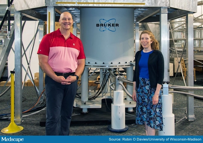
To reach the summit of two multimillion-dollar pieces of state-of-the-art equipment, scientists climb stairways spiraling around the structures, each the size of two supersized stacked refrigerators.
The $40 million National Science Foundation investment is intended, in part, to advance health research and drug development.
The spectrometers operate in much the same way as MRI scanners, the MRI machines used to take pictures to glimpse inside the human body. But instead of taking pictures of people, the new machines will take pictures of molecules, explains Jeffrey Hoch, PhD, from the Department of Molecular Biology and Biophysics at the University of Connecticut School of Medicine in Farmington.
Nuclear imaging will enable the study of molecules, atom by atom, and check chemical reactions under various conditions. The bigger the magnet in the machine, the finer the detail it can investigate.
The technology will help researchers understand battery components, nanomaterials, and surface coatings, and will open up myriad avenues of research, some yet to be imagined.

Prof Chad Rienstra (left), Prof Katherine Henzler-Wildman (right)
In less than 3 years, the University of Georgia in Athens and the University of Wisconsin at Madison will each have a cutting-edge 1.1 GHz spectrometer, and will join the UConn School of Medicine to make up the three pillars of the Network for Advanced Nuclear Magnetic Resonance. Researchers in Georgia will study substance mixtures and those in Wisconsin will study solids.
To use a spectrometer, someone climbs stairs wrapped around the machine and drops small sample-containing tubes into the top. “An air elevator” then carries them down into the magnet, where molecules can be isolated and studied, explains Engin Serpersu, PhD, a program director at NSF.
US Lags Behind Europe
There are only a handful of the spectrometers, which can cost up to $30 million each, in the United States, and outside researchers are rarely allowed access. So the addition of these two new machines will improve research capabilities considerably, says Steven Ellis, PhD, a program director at NSF.
This is good news because the United States has lagged behind Europe in ordering, installing, and using this technology, he says. In fact, that lag was noted in a 2013 National Research Council report that stressed the need for ultra-high-field nuclear imaging.
If the failure to keep up with advances in commercial technology “continues, the United States will probably lose its leadership role, as scientific problems of greater complexity and impact are solved elsewhere,” the report states.
“I can’t [overstate] the importance of making these instruments available to more users,” Ellis says. “If you want to know how a protein works, you really want to know how it’s folded, where all the atoms are, and how things are interacting with it.”
For the first time, the technology will be available to science, technology, engineering, and mathematics (STEM) students, primarily undergraduate institutions, minority-serving institutions and historically Black colleges and universities, and “any type of institution that can’t afford their own system but could prepare samples and use the data,” he explains. “It’s democratizing the technology.”
The NSF award goes beyond the spectrometers; it extends to cyber infrastructure, which includes the processing, storage, and sharing of data. It also covers the development of protocols so that people can use the knowledge bases to become experts.
The higher-field instruments speed up the collection of data, which is important because biologic samples are not always stable, Serpersu points out. Additionally, researchers can see how fast a single atom is moving, and “you can look at thousands of them simultaneously with NMR” or isolate some to study individually.
Potential Clues for Alzheimer’s and COVID
The technology could enhance study of the way proteins aggregate to cause neurologic diseases, such as Alzheimer’s, Serpersu says.
It could also advance research into antivirals for diseases like COVID-19, Ellis adds.
“If you want to interfere with spike-protein binding, it helps if you understand the structure of that and the structure of the receptor on the cell it binds to. Understanding those receptor structures can be very hard because they don’t crystallize well. Nuclear magnetic resonance is a better approach,” he says.
The Network for Advanced Nuclear Magnetic Resonance is starting with the three currently designated sites, but the expectation is that other centers will join the network and share resources and data, Ellis said.
The $40 million award does not cover the long-term costs of the program, so researchers will have to obtain grants to cover costs when they reserve time with the spectrometers. “The whole idea is to enable them to be more competitive by working on modern instrumentation and succeed in grant competitions,” Ellis says.
Marcia Frellick is a freelance journalist based in Chicago. She has previously written for the Chicago Tribune, Science News, and Nurse.com, and was an editor at the Chicago Sun-Times, the Cincinnati Enquirer, and the St. Cloud (Minnesota) Times. Follow her on Twitter at @mfrellick.
For more news, follow Medscape on Facebook, Twitter, Instagram, and YouTube
Source: Read Full Article
