
The severe acute respiratory syndrome coronavirus 2 (SARS-CoV-2) Omicron variant has shown significantly higher transmission rates than other variants, despite lower rates of severe disease. Researchers from Harvard Medical School are investigating the membrane fusion activity and structure of the spike protein of this variant in order to better understand these differences.
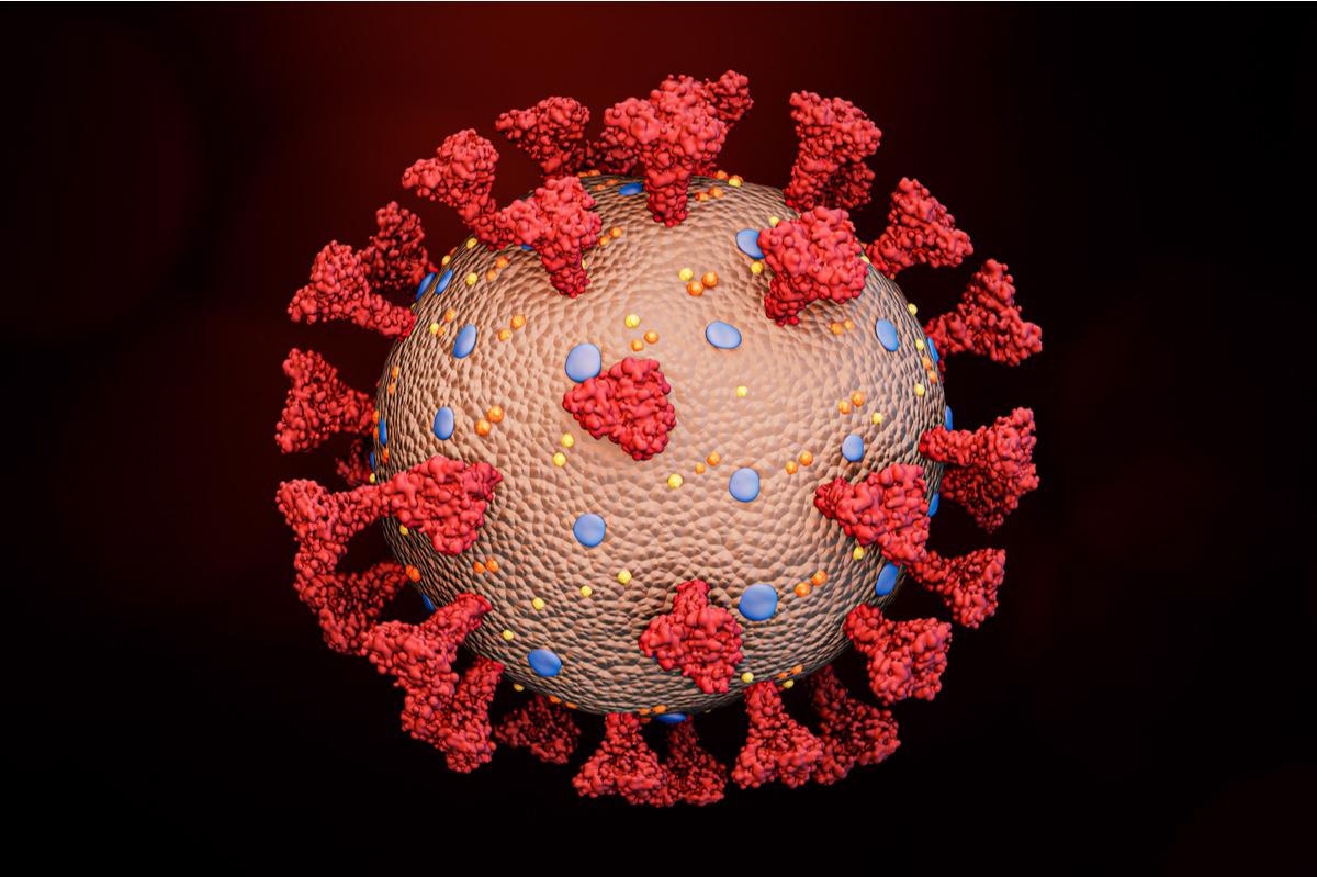 Study: Structural and functional impact by SARS-CoV-2 Omicron spike mutations. Image Credit: MattLphotography/Shutterstock
The researchers’ study can be found on the bioRxiv* preprint server, whilst the article undergoes peer review.
The Study
The researchers transfected HEK cells with an Omicron spike protein expression construct and compared the membrane fusion activity with constructs from variants such as Alpha, Beta and delta in order to characterize the full-length spike protein. All of the spike proteins were expressed at similar levels, although the Omicron proteins cleaved less between the two subunits at 24 hours post-transfection, which could suggest that the two mutations near the furin cleavage site do not enhance spike protein processing. Cells producing these proteins did fuse successfully with ACE2 cells, although the fusion activity of the Omicron spike protein was again lower than the other variants.
The scientists then carried out a time-course experiment with a cell-cell fusion assay, using both the spike protein and ACE2 at saturating levels, in order to better test whether the Omicron spike protein could induce membrane fusion more efficiently than other variants. Once again, there were no significant differences in fusion activity other than the Omicron spike protein showing slightly lower activity. When using cells expressing a minimum level of endogenous ACE2, all other variants showed significant fusion activities at later time points, but the Omicron spike protein was mostly inactive.
The researchers assumed that Omicron struggled to infect cells with low levels of ACE2, and so tried using HEK cells transfected with various amounts of ACE2. Once again, the Omicron spike protein lagged behind other variants, requiring a 10-fold increase in ACE2 in order to reach similar fusion activity. This was observed again when spike-producing cells were cotransfected with a furin expression construct, and the target cells were co-transfected with TMPRSS2.
The full-length Omicron spike protein was produced without any modifications by using a C-terminal strep-tagged construct for expression. The purified protein was then examined with gel-filtration chromatography. The wild-type Wuhan-Hu-1 spike protein is resolved into three peaks, which correspond to the prefusion trimer, and the postfusion S2 trimer and dissociated S1 monomer. Another variant, G614, shows a spike protein with a single peak of prefusion trimer.
The Omicron protein similarly presents one major peak, but there is a significant amount of aggregate on the leading side, and a shoulder on the trailing side. This suggests that the Omicron spike protein is significantly less stable than the G614 protein. Further analysis using SDS-PAGE reveals a large portion of uncleaved protein at 84 hours post-transfection. This supports the theory that Omicron struggles to transfect cells with low levels of ACE2 due to reduced cleavage of the spike protein.
Following this, the researchers attempted to determine the structure of the trimer based on cryo-EM images. 3D classification revealed three different classes for the spike protein, representing a closed position with all three receptor-binding domains (RBDs) facing ‘down’, a ‘one-RBD-up’ conformation and a RBD-intermediate conformation that has been seen in the G614 trimer. These were then further refined and modelled to reveal no major differences in the architecture of the full-length Omicron spike protein and the G614 spike protein. For both variants, the closed, all-down confirmation showed the N-terminal domain, RBD, C-terminal domain 1 (CTD1) and C-terminal domain 2 (CTD2) of S1 wrap around the S2 trimer. The one RBD up conformation preserved the helical core structure of S2 despite the RBD flipping up, which shifts the two adjacent N-terminal domains away and opens up the trimer significantly.
The researchers suggest that additional residues near the furin cleavage site that become significantly more structured due to the N679K mutation could reduce flexibility in the cleavage site, slowing the docking into the furin active site.
The Conclusion
The authors show that the Omicron spike protein requires significantly higher levels of ACE2 for efficient membrane fusion, lagging behind other variants. They also provided strong evidence that the Omicron spike protein is significantly less likely to be cleaved than other variants. With further investigation into the structure of the spike protein, they identify a mutation and resulting structural change that could be responsible for this change. This information could be key for drug developers, and could help identify the furin cleavage site as a target for treatment, potentially to alleviate the severity of disease.
Important notice:
bioRxiv publishes preliminary scientific reports that are not peer-reviewed and, therefore, should not be regarded as conclusive, guide clinical practice/health-related behavior, or treated as established information.
- Jun Zhang, et al. (2022). Structural and functional impact by SARS-CoV-2 Omicron spike mutations. bioRxiv. doi: https://doi.org/10.1101/2022.01.11.475922 https://www.biorxiv.org/content/10.1101/2022.01.11.475922v1
Posted in: Medical Science News | Medical Research News | Disease/Infection News
Tags: ACE2, Assay, Cell, Chromatography, Coronavirus, Coronavirus Disease COVID-19, covid-19, Medical School, Membrane, Mutation, Omicron, Protein, Protein Expression, Receptor, Research, Spike Protein, Transfection

Written by
Sam Hancock
Sam completed his MSci in Genetics at the University of Nottingham in 2019, fuelled initially by an interest in genetic ageing. As part of his degree, he also investigated the role of rnh genes in originless replication in archaea.
Source: Read Full Article